Introduction
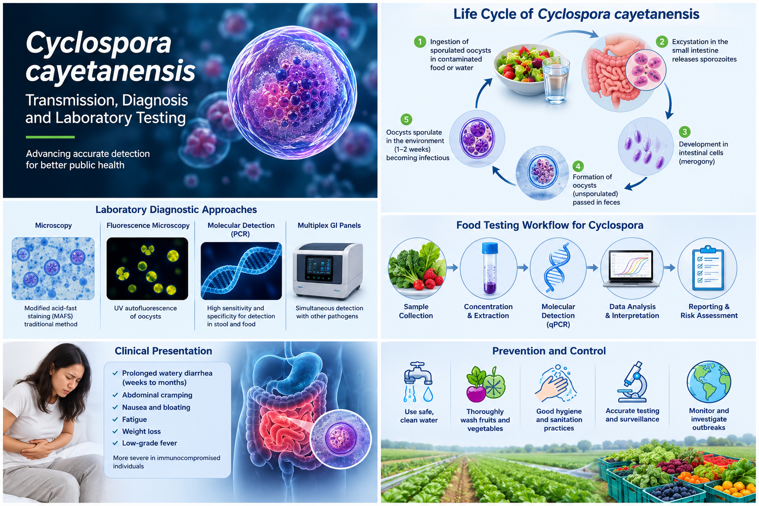
Cyclosporiasis is an intestinal infection caused by the human-specific protozoan parasite Cyclospora cayetanensis. Although North American outbreaks have been recognized since the 1990s, the parasite remains difficult to detect, trace and control because it is commonly present at low concentrations in food, water and clinical specimens. C. cayatanensis is endemic in tropical and subtropical regions including Nepal, Haiti, Mexico, Puerto Rico, Guatemala, and Peru [1].
A 2022 systematic review estimated the pooled prevalence of C. cayetanensis in water at 6.9%, with subgroup estimates of 3.46% for North America and 17.12% for irrigation water. These results should be interpreted cautiously because the studies used diverse concentration and detection methods and showed substantial heterogeneity. They support the need for standardized environmental surveillance, but they do not by themselves prove that the parasite's geographic range has shifted [2].
Transmission, incubation and clinical presentation
People become infected after ingesting food or water containing sporulated oocysts. Oocysts shed in human feces are unsporulated and are not immediately infectious; environmental maturation is required before transmission can occur. This biological delay makes immediate person-to-person transmission unlikely [3]. Most common outbreaks have been linked with contaminated fresh produce including basil, parsley, snow peas, raspberries, cilantro, mango, garlic, carrots, cantaloupe, cauliflowers, berries, salads, lettuce, etc.
Symptoms generally begin about one week after exposure, although onset can range from approximately 2 days to 2 weeks or longer. The most characteristic feature is explosive diarrhea without vomiting. Other symptoms include loss of appetite, abdominal cramping, bloating, increased gas, nausea, fatigue and weight loss. Without treatment, illness may persist for a month or longer and may relapse [4,5].
Trimethoprim-sulfamethoxazole is the treatment of choice. For immunocompetent adults, CDC describes a typical course of 160 mg trimethoprim plus 800 mg sulfamethoxazole twice daily for 7 to 10 days. No highly effective alternative has been established for patients who cannot take this medication; treatment decisions must therefore be individualized by a qualified clinician [6].
The 2026 U.S. outbreak
On July 24, 2026, CDC reported an open multistate outbreak investigation linked to iceberg lettuce. The notice listed at least 1,947 cases, 98 hospitalizations and no deaths across nine states. Taylor Farms de Mexico had recalled iceberg lettuce sourced from central Mexico, and the recalled product had been distributed in at least 27 states. Because outbreak counts and distribution information can change, readers should consult the current CDC and FDA notices before acting on these figures [4].
The outbreak illustrates why rapid communication among clinical laboratories, public-health agencies, food-testing laboratories, regulators and produce suppliers is essential. Patients may not seek care, routine stool testing may not include Cyclospora, and the implicated food may no longer be available by the time epidemiologic traceback identifies a likely source.
Why laboratory diagnosis is challenging
Clinical diagnosis is often delayed because Cyclospora testing may need to be requested specifically. Not all ova-and-parasite examinations or multiplex gastrointestinal panels include the organism. Oocysts may also be shed intermittently and at low levels, so a single negative specimen does not exclude infection. CDC advises that several stool specimens collected on different days may be needed when suspicion remains high [4].
Concentration procedures improve recovery. Microscopy can use wet mounts, modified acid-fast staining, modified hot-safranin staining, differential-interference contrast and ultraviolet fluorescence. Oocysts are approximately 8–10 micrometres in diameter and characteristically autofluoresce. Modified acid-fast staining is variable, so size, morphology and fluorescence should be interpreted together [4].
Molecular assays can improve analytical sensitivity and reduce dependence on microscopic expertise. Varma and colleagues developed an 18S rRNA real-time PCR assay capable of detecting DNA from as few as one oocyst per reaction under the study conditions [7]. However, high sensitivity must be balanced against specificity, particularly when conserved targets are shared with related coccidia. Scientist reported primers and probes cross react with Eimeria mitis and Toxoplasma gondii [8].
Food and environmental testing: sensitivity versus specificity
Detecting C. cayetanensis in produce or water is more complex than testing stool. Oocysts must first be recovered from a large and heterogeneous matrix, concentrated, disrupted and subjected to nucleic-acid extraction. Low organism burden, uneven contamination and PCR inhibitors can all reduce detection.
The U.S. FDA Bacteriological Analytical Manual Chapter 19b has historically used an 18S rRNA real-time PCR method. Independent verification by the Canadian Food Inspection Agency showed that assay exclusivity could change with the qPCR master mix: three of five mixes tested were exclusive for C. cayetanensis, while two also amplified several Eimeria species and Isospora suis. The study therefore recommended local exclusivity testing when laboratories change reagents, instruments or conditions [9].
The same verification study highlighted a limitation of confirmatory sequencing for short 18S amplicons. Many sequencing reactions failed, assembled reads were only 50–60 base pairs, and the region was conserved rather than unique to C. cayetanensis. A short sequencing result may therefore add little diagnostic discrimination, even when it matches the expected target [9].
To improve specificity, Durigan and colleagues developed mitochondrial markers, including the Mit1C target within the cox3 region. The optimized Mit1C assay was designed to reduce cross-reactivity while retaining useful sensitivity for produce and agricultural-water testing [8]. A 13-laboratory validation subsequently found statistically similar detection performance for Mit1C and the reference 18S assay [10]. At a five-oocyst inoculum in romaine lettuce, overall detection was 69.23% with Mit1C and 61.54% with 18S; Mit1C specificity was 98.9% [10].
The May 2026 FDA BAM Chapter 19b edition uses the Mit1C approach and includes an approved matrix extension for fresh basil and parsle [11]. The FDA’s current method (BAM 19b) is optimized strictly using the commercial PrimeTime Gene Expression Master Mix and was originally validated on the ABI7500 with 40cycles of 95C for 15s and 67C for 1 min following an initial denaturation step of 95C for 3min [11]. A 2026 evaluation detected as few as five oocysts in basil, parsley and broccoli, but low-level detection was less frequent in broccoli, likely because the florets reduced wash recovery. This reinforces the need for matrix-specific validation rather than assuming that one food type predicts performance in another [12].
Genomic similarity and the need for careful assay design
Assay design is complicated by the close evolutionary relationship between Cyclospora and Eimeria. Comparative analysis showed that the apicoplast and mitochondrial genomes of C. cayetanensis and Eimeria tenella share approximately 85.6% and 90.4% nucleotide identity, respectively, with conserved gene organization [12]. The nuclear genome is approximately 44 megabases, while the mitochondrial genome is approximately 6.2 kilobases [12,13].
This similarity does not make specific detection impossible, but it means that primer and probe design must be assessed against current sequence databases and a relevant exclusivity panel. Validation should include related coccidia, alternate master mixes, multiple instruments and realistic food matrices. Laboratories should also monitor internal amplification controls to identify inhibition. Lab validation and verification should independently verify the assay on different equipment, using different reagents and test all species of Eimeria and Toxoplasma to confirm level of cross reactivity.
Genomics and outbreak investigation
Whole-genome and targeted sequencing offer opportunities for linking clinical, food and environmental samples, but low template concentration remains a major obstacle. Enrichment strategies are therefore more practical than untargeted whole-genome sequencing for many produce samples.
A targeted amplicon sequencing method developed by Leonard and colleagues recovered at least 24 markers from leafy greens inoculated with as few as 10 oocysts and successfully genotyped clinical specimens with low parasite burden. Such approaches can support genetic clustering, source tracking and outbreak investigation while using far less DNA than conventional whole-genome sequencing [15].
Canadian surveillance data also show why typing capacity matters. From 2000 to 2022, 5,337 cyclosporiasis cases were reported in Canada, with seasonal peaks from May to August and increasing incidence over time. International travel was a common exposure, while national investigations frequently identified produce items of interest but rarely confirmed a single source. The authors emphasized real-time genetic typing as an important future capability [16].
Practical priorities for laboratory validation
A laboratory introducing or modifying a Cyclospora assay should consider the following priorities:
Verify analytical sensitivity across realistic low oocyst concentrations rather than relying only on purified DNA.
Evaluate inclusivity and exclusivity using current sequence databases and DNA from related coccidia.
Assess the effect of master-mix lots, reagent substitutions, thermal-cycling conditions and instrument platforms.
Test multiple produce matrices because wash recovery, inhibition and detection can vary substantially by commodity.
Include extraction controls, no-template controls, positive controls and internal amplification controls.
Define repeat-testing and confirmation rules before implementation.
Avoid assuming that a short conserved amplicon provides definitive sequencing confirmation.
Document all deviations from the validated method and perform bridging studies before routine use.
Conclusion
Cyclospora cayetanensis remains a challenging organism for clinical, food and environmental laboratories. The central problem is not simply whether PCR can detect the parasite; it is whether the complete workflow can recover low numbers of oocysts, remove inhibitors and distinguish C. cayetanensis from closely related organisms under real-world conditions.
The transition from conserved 18S rRNA assays toward more specific mitochondrial targets is an important advance, but it does not eliminate the need for local verification. Matrix effects, reagent chemistry, instrument settings and confirmation strategies can materially change assay performance. Future outbreak response will benefit from combining validated qPCR with sensitive targeted sequencing methods capable of linking low-burden clinical and food specimens.
References
Dixon BR, B. J. (2005). Detection of Cyclospora cayetanensis oocysts in human fecal specimens by flow cytometry. J Clin Microbiol., 43(5):2375-9.
Naganathan T, O’Connor A, Sargeant JM, Shapiro K, Totton S, Winder C, Greer AL. The prevalence of Cyclospora cayetanensis in water: a systematic review and meta-analysis. Epidemiol Infect. 2022;150:e15. doi:10.1017/S0950268821002521. https://doi.org/10.1017/S0950268821002521
Ortega YR, Sanchez R. Update on Cyclospora cayetanensis, a food-borne and waterborne parasite. Clin Microbiol Rev. 2010;23(1):218-234. doi:10.1128/CMR.00026-09. https://doi.org/10.1128/CMR.00026-09
Centers for Disease Control and Prevention. Clinical overview of cyclosporiasis. March 4, 2024. https://www.cdc.gov/cyclosporiasis/hcp/clinical-overview/index.html
Centers for Disease Control and Prevention. Cyclospora outbreak linked to iceberg lettuce in 9 states. July 24, 2026. https://www.cdc.gov/cyclosporiasis/outbreaks/07-26/index.html
Centers for Disease Control and Prevention. Clinical care of cyclosporiasis. March 8, 2024. https://www.cdc.gov/cyclosporiasis/hcp/clinical-care/index.html
Varma M, Hester JD, Schaefer FW 3rd, Ware MW, Lindquist HDA. Detection of Cyclospora cayetanensis using a quantitative real-time PCR assay. J Microbiol Methods. 2003;53(1):27-36. doi:10.1016/S0167-7012(02)00209-9. https://doi.org/10.1016/S0167-7012(02)00209-9
Durigan M, Ewing-Peeples L, Almeria S, Balan KV, Grocholl J, Irizawa S, Mammel M. Detection of Cyclospora cayetanensis in food and water samples: optimized protocols for specific and sensitive molecular methods from a regulatory agency perspective. J Food Prot. 2024;87(7):100291. doi:10.1016/j.jfp.2024.100291. https://doi.org/10.1016/j.jfp.2024.100291
Lalonde L, Oakley J, Fries P. Verification and use of the US-FDA BAM 19b method for detection of Cyclospora cayetanensis in a survey of fresh produce by CFIA laboratory. Microorganisms. 2022;10(3):559. doi:10.3390/microorganisms10030559. https://doi.org/10.3390/microorganisms10030559
Almeria S, Grocholl J, Mullins J, et al. Multi-laboratory validation of a modified real-time PCR assay (Mit1C) for the detection of Cyclospora cayetanensis in fresh produce. Food Microbiol. 2025;128:104727. doi:10.1016/j.fm.2025.104727. https://doi.org/10.1016/j.fm.2025.104727
U.S. Food and Drug Administration. BAM Chapter 19b: Molecular detection of Cyclospora cayetanensis in fresh produce using real-time PCR. May 2026 edition. https://www.fda.gov/food/laboratory-methods-food/bam-chapter-19b-molecular-detection-cyclospora-cayetanensis-fresh-produce-using-real-time-pcr
Rogers E, Almeria S. Validating the updated BAM Chapter 19b method for the detection of Cyclospora cayetanensis in outbreak-linked fresh produce. J Food Prot. 2026;89(6):100782. doi:10.1016/j.jfp.2026.100782. https://doi.org/10.1016/j.jfp.2026.100782
Tang K, Guo Y, Zhang L, et al. Genetic similarities between Cyclospora cayetanensis and cecum-infecting avian Eimeria spp. in apicoplast and mitochondrial genomes. Parasit Vectors. 2015;8:358. doi:10.1186/s13071-015-0966-3. https://doi.org/10.1186/s13071-015-0966-3
Liu S, Wang L, Zheng H, et al. Comparative genomics reveals Cyclospora cayetanensis possesses coccidia-like metabolism and invasion components but unique surface antigens. BMC Genomics. 2016;17:316. doi:10.1186/s12864-016-2632-3. https://doi.org/10.1186/s12864-016-2632-3
Leonard SR, Mammel MK, Gharizadeh B, et al. Development of a targeted amplicon sequencing method for genotyping Cyclospora cayetanensis from fresh produce and clinical samples with enhanced genomic resolution and sensitivity. Front Microbiol. 2023;14:1212863. doi:10.3389/fmicb.2023.1212863. https://doi.org/10.3389/fmicb.2023.1212863
Morton V, Janicki R, Dumoulin D, Dixon B, Guy R. Epidemiology of Cyclospora cayetanensis infections in Canada: 2000-2022. Foodborne Pathog Dis. 2025. doi:10.1177/15353141251377318. https://doi.org/10.1177/15353141251377318
Disclaimer: This article is intended for scientific and educational purposes. It does not replace validated laboratory procedures, regulatory guidance or individual medical advice.